
This video is part of the blood pressure regulation series. We discuss the baroreceptors and their contribution for maintaining the blood pressure. We will discuss the various type of mechanisms that help control the blood pressure. {article:https://articles.drbeen.com/2016/10/19/blood-pressure-control-by-baroreceptors/}
CVS PHYSIOLOGY LECTURE # 16 STUDY NOTES:
BLOOD PRESSURE CONTROL BY BARORECEPTORS
The MAP i.e. mean arterial pressure, also considered as the perfusion pressure, is taken as the pressure difference between the arteries and the veins. The regulation of blood pressure is done in order to maintain the MAP. The MAP hence dictates the amount of oxygen and nutrients that is supplied by the blood vessels and the waste that is carried away from the tissues.
The body has the ability to counteract long term as well as short term changes in blood pressure. The long term pressure changes cause the body to respond through the activation of renin-angiotensin system.
Rapid/short term changes in blood pressure compel the body to activate the following receptors:
• Baroreceptor present on the arch of aorta and carotid sinus
• Chemoreceptors present all over the body, aorta and carotid sinuses
• Atrial receptors present on the wall of right atrium
These receptors are modified nerve endings that are sensitive to rapid offsets in blood pressure. Rapid offsets in pressure can occur, for example, in a previously standing person who suddenly sits down. During the process, a large volume of blood is shifted from the peripheral to the central regions of the body. Consequently, a large volume of blood enters the heart and this volume overload (increased preload) causes the heart to increase its cardiac output. A simultaneous increase in blood pressure will also be observed with increase in cardiac output. The increase in blood pressure is registered by the baroreceptors which are densely situated on the walls of the arch of aorta and the carotid sinus which is present on internal carotid artery.
Similarly, a drop in blood pressure is registered by the baroreceptors when the person stands up suddenly from a sitting position. These pressure sensing bodies are modified nerves with stretch receptors on their ends. These stretch receptors are attached to the cytoskeleton present within the nerve endings. These nerve endings are called spray type nerves. High blood pressure in the blood vessels causes stretch of these receptors which results in movement of sodium ions into the nerve endings, thereby, initiating an action potential.
These baroreceptors have a baseline firing pattern. That means they have an intrinsic potential to generate action potentials at a particular frequency at all times. This frequency is increased when the baroreceptors receive a stretch stimulus secondary to increase in blood pressure. The carotid sinuses increase their rate of impulse generation when the pressure in them builds up to values greater than 50 mm Hg. Below thisthreshold pressure, the carotid baroreceptors fail to initiate an action potential. On the other hand, the arch of aorta can record drops in blood pressure up to 30 mm Hg. The upper limit for blood pressure, after which the frequency of action potential stops increasing, is 175 mm Hg. The normal MAP is calculated to be 93 mm Hg. At this pressure, the baroreceptors are believed to be the most sensitive and even slight changes in pressure will result in rapid firing of action potentials. At blood pressures lower than 30 mm Hg, the chemoreceptors come into play. The chemoreceptors function by sensing the arterial concentration of carbon dioxide, oxygen, Ph and other metabolites rather than detecting changes in blood pressure.
STRUCTURE OF THE CAROTID SINUS
The carotid sinus is present on the base of internal carotid artery at the level of bifurcation of the common carotid artery. The sinus area is slightly dilated as the tunica media which is normally comprised of muscles, is relatively thin. The tunica adventitia, on the other hand, is thicker than usual. This is the layer of the blood vessels where the nerve receptors are situated. Same is true for the location of baroreceptors on the arch of aorta.
THE BARORECEPTOR REFLEX
The baroreceptor reflex, like other reflex arcs, is comprised of three units:
1) Afferent nerve carrying impulses from the receptors,
2) Central processing unit
3) An efferent nerve that innervates the effector
Afferent impulses from the carotid sinus are carried by the Herring nerve, a branch of Glossopharyngeal nerve (CN-9). In the case of baroreceptors present on the arch of aorta, the Vagus nerve (CN-10) is the afferent nerve that carries impulses to the spinal cord. Both, the Vagus nerve and the Glossopharyngeal nerve, feed impulses from the baroreceptors into the nucleus of tractus solitarius. These nuclei are situated in the medulla of the spinal cord and their job is to process the incoming afferent impulses. Also within the Medulla and lower 1/3rd of the Pons, there arevasoconstricting center, the vasodilatory center and the cardio-inhibitory center. These centers receive processed impulses from the nucleus of tractus solitarius and from here efferent impulses in the form of sympathetic and parasympathetic nerves arise. Impulses are carried to the heart via the parasympathetic Vagus nerve. Sympathetic impulses travel down the intermedio-lateral segment of the spinal cord and give rise to efferent motor spinal nerves which enter the sympathetic ganglion running parallel to the spinal cord. Postganglionic sympathetic nerves ultimately supply the heart and the peripheral vasculature. Another preganglionic sympathetic nerve also supplies the adrenal medulla which results in the release of epinephrine and norepinephrine, which further contribute in enhancing the sympathetic activity. The end result is either an increase or decrease in the blood pressure, thereby correcting the disturbance in hemodynamics of the body. This phenomenon is also referredto as the buffering effect, since the change in pressure is buffered back to normal. The Vagus and Glossopharyngeal nerves, because of the same reason, are therefore known as the buffering nerves.
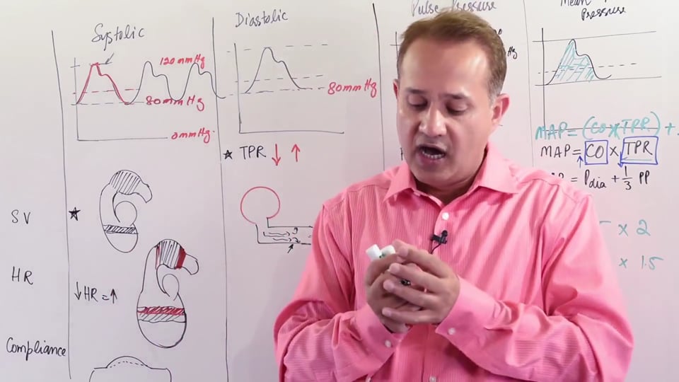
The factors responsible for change in mean arterial pressure are formulated as follows:
➢ MAP = Heart Rate x Cardiac Output (CO)
Whereas, CO = SV (stroke volume) x TPR (total peripheral resistance)
Therefore, MAP = HR x SV x TPR
The stroke volume is altered by altering the force of contractility of the heart muscles. The sympathetic nerves supplying the heart muscles affect the stroke volume. The parasympathetic nerves supplying the SA and AV node are responsible for producing changes in heart rate. The TPR can be increased or decreased by changing the diameter of peripheral vasculature which is under the control of the sympathetic nervous system.
EFFECTS OF BARORECEPTORS DURING VARIOUS CONDITIONS
1) DUE TO CHANGES IN BLOOD PRESSURE
a. Reduced Blood Pressure: Reduction in blood pressure will result in a decrease in the number of afferent impulses from the baroreceptors. The sympathetic activity will increase and as a result, the TPR, HR and the stroke volume will allincrease. At the same time, the parasympathetic input will taper down. All these changes will result in increasing the blood pressure back to normal
b. Increased Blood Pressure: This happens in situations like exercise or stress. Increased blood pressure will result in stretching of the stretch receptors. This increases the frequency of afferent impulses. Sympathetic supply will decrease and the parasympathetic system will take over. Finally, the blood pressure is decreased back to normal.
2) DUE TO CHANGES IN CARDIAC OUTPUT
a. Decreased Cardiac Output: Occurs in situations of vomiting, diarrhea, hemorrhage etc. As a result of these, both the volume, and therefore pressure of the blood decreases. Afferent impulse firing of the baroreceptors decreases. As a consequence, there’s a sympathetic overflow which causes an increase in HR, TPR and SV. Due to an increase in these parameters, the blood pressure is raised back to normal.
b. Increased Cardiac Output: There’s an increased impulse generation from the baroreceptors due stretch caused by increased volume of blood. This increased afferent input from the baroreceptors results in activation of the PANS. Once activated, the parasympathetic nervous system decreases the blood pressure back to normal.
3) MASSAGING THE CAROTID SINUS
Massaging the carotid sinuses physically increases the pressure on the baroreceptors present there. The carotid baroreceptors respond by increasing the rate of afferent impulse firing. The sympathetic system will be shut down and the parasympathetic system is activated. This results in events leading to decrease in blood pressure of the body. Patients who have atrial arrhythmias are asked to massage carotid sinus to decrease heart rate.
4) STENOSIS OF CAROTIDS
Stenosis of carotids proximal to the sinus or obstruction of the carotids due to atherosclerosis will cause the baroreceptors to register a decrease in pressure. Therefore, sympathetic system activation follows. Increased sympathetic activity causes a resultant increase in blood pressure. This increase in blood pressure may cause hypertension in an otherwise normal person.
The above factors and their affect on the baroreceptor response are summarized in the table below:
|
FACTOR |
AFFERENT |
SANS ACTIVITY |
PANS ACTIVITY |
BLOOD PRESSURE |
HEART RATE |
VASCULAR RESPONSE |
|
↓BP |
↓ |
↑ |
↓ |
↑ towards normal |
↑ |
Vasoconstriction |
|
↑BP |
↑ |
↓ |
↑ |
↓ towards normal |
↓ |
Vasodilation |
|
↓CO |
↓ |
↑ |
↓ |
↑ towards normal |
↑ |
Vasoconstriction |
|
↑CO |
↑ |
↓ |
↑ |
↓ towards normal |
↓ |
Vasodilation |
|
CAROTID MASSAGE |
↑ |
↓ |
↑ |
↓ towards normal |
↓ |
Vasodilation |
|
CAROTID STENOSIS |
↓ |
↑ |
↓ |
↑ towards normal |
↑ |
Vasoconstriction |
It’s important to understand that baroreceptor control of BP is a short term regulation of blood pressure. Any short term derangements are dealt via the baroreceptor response, whereas long term control of the BP is controlled via the RAAS (Renin Angiotensin Aldosterone System). The baroreceptors alsohave the ability to adapt to chronic changes in blood pressure. If the mean pressure is changed over time to a new value, the baroreceptors will start using that MAP as the baseline. Any subsequent blood pressure changes will then be rectified keeping in view the new baseline value of MAP.
In this video we will learn about :
1. Short term regulation mechanisms of blood pressure.
2. Location of baroreceptors.
3. Structure of stretch baroreceptors.
4. Structure of carotid sinus.
5. Afferent and efferent fibres of baroreceptors.
6. Regulation of increased blood pressure by baroreceptors.
7. Regulation of decreased blood pressure.
No credit card information needed.


Write A New Comment
0 Comments